 By
By
- Davor Ježek M.D., Ph.D., Department of Histology and Embryology, University of Zagreb, Zagreb, Croatia
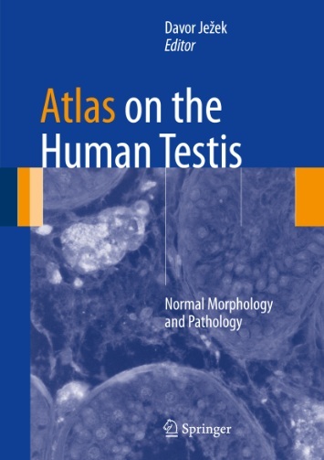
Atlas on the Human Testis: Normal Morphology and Pathology presents histological illustrative material from paraffin and semi-thin sections of the human testis which are routinely used in the assessment of testicular morphology, allowing an early detection of carcinoma in situ and more advanced pathological changes of the testicular parenchyma. The early detection of cancer in situis based on the careful morphological investigation of the biopsy and immunohistochemistry (IHC). Therefore, this atlas contains detailed descriptions of IHC methods as well as modern molecular biological methods such as DNA microarrays and proteomics and advanced microscopy techniques related to the testicular biopsy.
Adequate evaluation of the testicular biopsy leads to high cure rates of testicular neoplasms which can be used as a basis to successfully treat infertility in men.
Atlas on the Human Testis: Normal Morphology and Pathology is a valuable reference tool which will appeal to andrologists, urologists, pathologists, clinical embryologists, as well as reproductive biology scientists.
Key Features
- Details richly illustrated normal spermatogenesis and various degrees of spermatogenesis disorders allowing the reader to estimate the chances of a successful spermatozoa extraction (TESE) despite the damaged spermatogenesis
- The Human Testis: Normal Morphology and Pathology contains detailed descriptions of seminoma and non-seminoma tumours providing the reader with a clear overview of the testicular neoplasms and their nature and prognosis
- Describes new methods for the analysis of the testicular biopsy familiarising the reader with new approaches of the analysis of the testicular tissue that could be routine methods in the future


